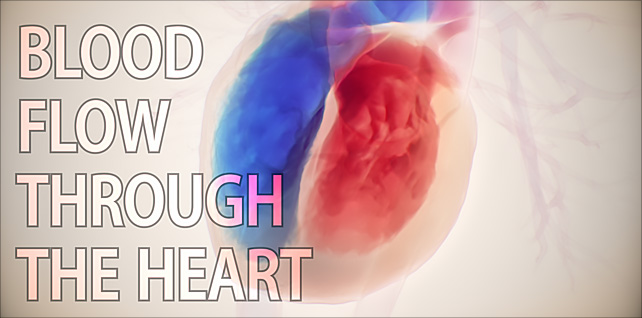
Blood flow through a beating heart | Cardiac hemodynamics
Sep 11, 2023The heart’s internal blood flow is complex, beautiful, and driven by several components, including the four heart valves, ventricular makeup, the rhythmic contraction and relaxation cycles, the automatic nervous system, and hormonal influence. All these factors work in tandem to ensure continuous and efficient circulation of blood throughout the heart and the entire body.
Our goal in this scene was to emulate blood flow in the chambers of the heart using fluid dynamics (in Houdini).
Simulations of this type require a high amount of detail in order to contend with the high velocities of pressure and flow, and to address the thin collision geometry in the valves and between the chambers.
We rendered this in a style that would feature the blood flow and give the heart more of a fluoroscopy look & feel. Oxygenated blood is shown in red; deoxygenated blood in blue.
The percentage of blood that is pumped out with each beat is called ejection fraction or EF. Normal ejection fraction is greater than 55%.
Comprehending the dynamics of blood flow is pivotal for grasping the nuances of cardiovascular physiology. Analyzing these flow patterns is instrumental in evaluating cardiac performance, specifically when assessing the heart’s diastolic function. Understanding the efficiency of the ventricles filling up before they contract can be crucial when diagnosing heart failure.
Tagged: 3D blood flow visualization, aorta, biology, biomechanics, blood flow, blood flow in heart, Blood flow inside the heart, blood pressure, cardiac anatomy, cardiac hemodynamics, cardiac physiology, cardiovascular system, circulatory system, contraction, ejection fraction, electrophysiology system, fluid mechanics, haemodynamics, heart failure, heart valves, heartbeat, hemodynamics, HFpEF, HFrEF, houdini, Houdini fx, hybrid medical, Intraventricular hemodynamics, med art, medical animation, pharma, preserved ejection fraction, science visuals, scientific animation, sidefx, stroke volume, ventricular volume
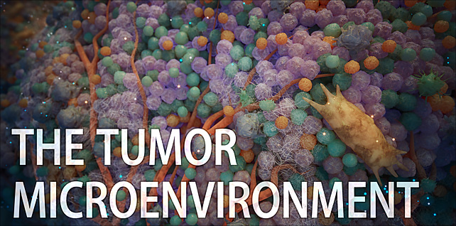
A closer look at the tumor microenvironment (TME)
Oct 27, 2023This animation delves deep into the tumor microenvironment (TME) — a complex cellular domain in which a tumor resides.
Take an immersive, cinematic journey through this thriving tumor microenvironment, showcasing the intricate biological landscape of an extracellular matrix, newly-forming blood vessels, cancer-associated fibroblasts, and various immune cells such as macrophages, T cells, B cells, dendritic cells, neutrophils, NK cells, and more.
Tumors are more than just masses of malignant cells. Although the composition of the TME varies between tumor types, in many cases, the actual tumor cells might comprise less than half of the tumor’s total mass. Our animation also highlights the newly-formed vasculature. These vessels not only sustain the TME with oxygen and nutrients but also provide a route for tumor cells to access the bloodstream and potentially metastasize to distant sites.
In this exploration, we also emphasize cell migration — a critical, multifaceted process shaped by various factors. Cells within the TME communicate with each other through an intricate network of signaling pathways that can influence immune response and surveillance, angiogenesis, tumor growth, metastasis, and resistance to therapies. There have been recent advancements in understanding the spatial arrangement and complex interrelationships of cells in the tumor microenvironment.
Ultimately, a tumor’s growth and potential to spread are shaped by several intertwined pathological events, including chronic inflammation, alterations and remodeling of the extracellular matrix, angiogenesis, immune system suppression, and cell migration.
Tagged: angiogenesis, bioart, biology, biomedical communications, biotech, cancer cells, cancer research, cell biology, cell migration, extracellular matrix, hybrid medical, immune cells, immuno oncology, immuno-oncology, immunotherapy, medical animation, medical visualization, metastasis, microscopy, oncology, pharma, science, science visuals, scientific animation, spatial biology, T cells, t lymphocytes, tumor, tumor heterogeneity, tumor microenvironment
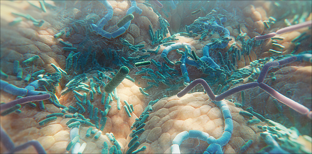
The diverse human gut microbiome
Jun 06, 2023Animation sequence featuring a fly-over of the human gut microbiome – a huge, diverse range of many different types of bacteria residing inside our small and large intestine.
This sequence was created for an episode (for WebMD) that examines how the gut microbiome may influence cancer response to immune checkpoint inhibitors. Essentially, if a patient has melanoma, the types of bacteria in their gut may help determine if the cancer responds to checkpoint inhibitor therapy or not.
There are trillions of microbes in our gastrointestinal tract, around 90% of which are bacteria.
Our microbiome has a profound impact on our well-being and can affect us in several different ways – both beneficial and harmful. In recent years, scientists and researchers have discovered correlations between the composition of an individual’s microbiome and their wider health, including obesity, mental health, bowel disease, and the immune system.
Further advances need to be made in learning more about actual causation – and which effects come from which individual microbes.
Tagged: bacteria, biology, biomedical communications, biotech, biotechnology, gastrointestinal tract, gut bacteria, gut diversity, gut health, gut microbes, gut microbiota, human gut microbiome, hybrid medical, medical animation, Medical science, medical visualization, medicine, microbiology, microbiome, microbiome health, microbiome medicine, pharma, science, science visuals, scientific animation, visual science
Oligomers, fibrils, & plaques | Alzheimer’s | medical animation
Aug 21, 2023Segment from 5-minute mechanism of action animation.
This sequence explores a hallmark and pathophysiological feature of Alzheimer’s disease: toxic amyloid beta aggregates (Aβ) that accumulate into plaques (neurofibrillary tangles) near neurons in the brain.
A challenge for this sequence was to show the assembly of these naturally occurring peptides – oligomers, fibrils, and plaques – all in one continuous shot.
In addition, we show how over time, extracellular amyloid beta aggregates, including oligomers, fibrils, and plaques, are thought to exert toxic effects on neurons and synapses, disrupt cell function, and lead to synaptic dysfunction and loss.
Tagged: aggregates, alzheimer’s, alzheimer's disease, alzheimers, amyloid, amyloid beta, amyloid beta plaques, amyloid fibril, amyloid plaques, biomedical communications, biotech, brain deterioration, cognitive decline, digital health, extracellular amyloid beta aggregates, fibrils, hybrid medical, medart, medical animation, medical visualization, neurofibrillary tangles, neuroscience, oligomers, plaques, scicomm, synaptic dysfunction, toxic aggregates
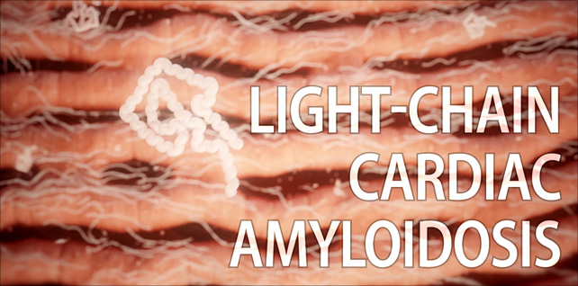
Amyloid light chain (AL) amyloidosis | cardiac amyloidosis animation
Jul 21, 2023Opening with a view of the heart, we reveal several amyloid deposits infiltrating the muscle tissue.
Then we zoom down to the heart muscle cells and see misfolded light chains aggregating into insoluble amyloid fibrils.
We fade, over time, from the early onset of light-chain infiltration to late-stage fibril aggregation, where the buildup of amyloid deposits around and in between the cardiomyocytes has made the muscle tissue stiff and less flexible, impairing the heart’s ability to relax and fill with blood.
Amyloid light chain (AL) amyloidosis is the most common form of systemic amyloidosis and is caused by the misfolding of immunoglobulin light chains, which are produced by abnormal plasma cells in the bone marrow.
Deposits can be widespread, affecting multiple organs, or they can be more localized.
Tagged: AL amyloidosis, amyloid deposits, amyloid light chain, amyloidosis, cardiac, cardiac amyloidosis, cardiology, fibrils, heart, heart failure, heart health, hybrid medical, immunoglobulin light chains, light chain amyloidosis, medical animation, misfolded proteins, misfolding, myocardium, scientific animation, visual science
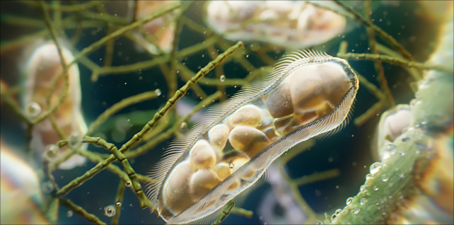
Paramecia: unicellular ciliates animation
Jun 02, 2023A view of paramecia spiraling their way about in a freshwater pond.
In this sequence, we get a close-up look at one of these unicellular microorganisms as it makes its way around using the tiny hair-like cilia on its outer surface. Arranged in longitudinal rows, there are around 4,000 of these motile cilia on its surface, beating in waves to ensure movement and feeding.
In addition to other organelles, we can see several vacuoles inside, digesting organisms such as bacteria and yeasts.
The paramecium was among the first single-celled organisms to be observed under the microscope.
In order to capture the emergent behavior and movement of this microworld, the definitive goal here was to create a natural environment using physics simulations and a procedural pipeline instead of hand-keyed animation.
Tagged: biology, cell, ciliate, ciliates, houdini, houdinifx, hybrid medical, medical animation, micro, microbes, microbiology, microcosmos, microscope, microscope photography, microscopy, microworld, paramecia, paramecium, pharma, photomicrography, redshift, redshift3d, science, science visuals, scientific animation, sidefx, unicellular
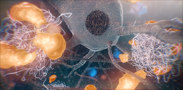
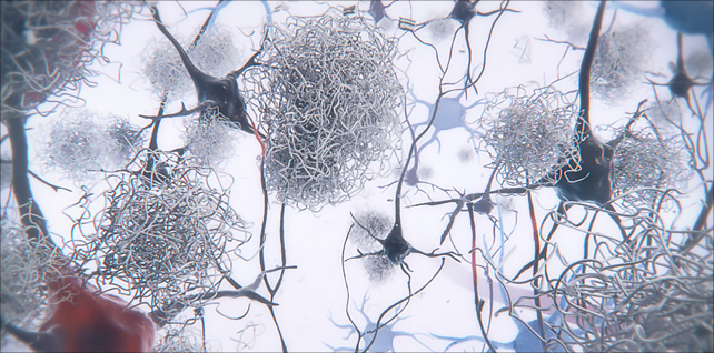
Proliferation and activation of microglia in the brain
Aug 31, 2023Amyloid plaques are the hallmark of Alzheimer’s disease (AD).
This sequence shows the proliferation and activation of microglia in the brain, concentrating around and engulfing neurotoxic amyloid-beta plaques that have caused damage to impaired neurons nearby.
Microglia are specialized immune cells of the central nervous system, specifically in the brain and spinal cord. They play a vital role in monitoring and maintaining the neural environment. One of their primary responsibilities is to detect and remove damaged neurons, plaques, and other cellular debris from the brain.
We worked closely with our clients to create an accurate portrayal of microglia, including their form and structure, their movement and behavior, as well as the manner in which they form a barrier around these growing, toxic plaques. Microglia size, movement, and morphology were all examined by viewing time-lapse imaging projection software – and then recreated in 3D.
One potentially harmful aspect of this relationship between microglia and plaques is that prolonged activation of microglia in response to persistent plaques can lead to chronic inflammation, which might contribute to neuronal damage and the progression of Alzheimer’s disease.
Tagged: aggregates, Alzheimer’s disease, alzheimers, amyloid, amyloid beta, amyloid fibril, amyloid plaques, amyloid-beta plaques, axonal dystrophy, Aβ plaques in Alzheimer’s disease, biomedical communications, biotech, biotechnology, brain deterioration, cognitive decline, glia, hybrid medical, medical animation, medical visualization, microglia, microglia barrier, microglial, microglial phagocytosis, Minneapolis Minnesota, nervous system, neurofibrillary tangles, neurons, neuroprotection, neurotoxic, plaques, scientific animation, synaptic dysfunction, toxic aggregates